Scientific Details
Family level detail.
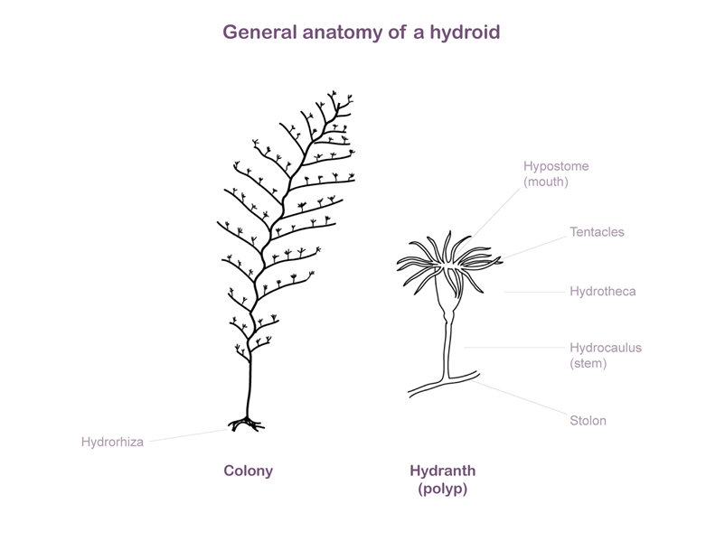
Erect branched or unbranched plumose stems. Stems and hydrocladia divided into segments (internodes). Hydrocladia alternate on stem internodes. Hydrotheca saccate, attached at base to hydrocladial internode, margin usually cuspate. Three nematothecae attached to each hydrotheca - one tubular median with a terminal orifice, fused to upper (anterior) side of hydrotheca and smaller twin laterals, one at each side of base of hydrotheca. Nematothecae similar to laterals on internodes of main stem. Gonophores fixed sporosacs containing eggs and sperm, protected by a gonotheca; accessory protective structure (corbula) in some families.
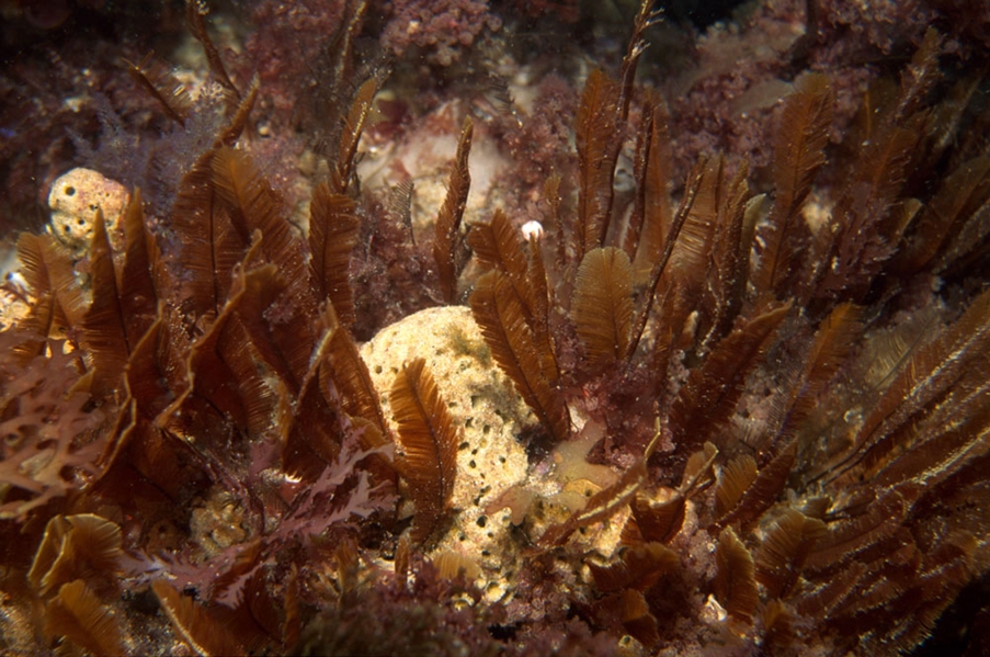
The most spectacular and graceful hydroids in southern Australia belong to the Aglaopheniidae. Preliminary field identification of many aglaophenian species can be made from observing colony size, structure and colour, however, precise identification usually requires microscopic examination. In the genus Aglaophenia the corbula is important in identification, including its size, the number and shape of leaflets (pinnae), whether the pinnae are free from one another (open corbula) or connected together by tissue (closed corbula), and the shape of the nematothecae on the pinnae. When fertile, species of the Aglaopheniidae can be identified from the presence, absence or development of the corbula.
Provisional identification is possible by plucking several hydrocladia from a specimen stem and laying them in a drop of water on a glass microscope slide, compressing them gently under a coverslip and examining them microscopically. The important structures of the hydrocladium, hydrotheca and nematotheca can usually be seen under low power magnification. Often however, the dense and strongly coloured internal tissue (coenosarc) obscures diagnostic structures. The coenosarc can be dissolved in domestic bleach (calcium hypocholorite solution) diluted in tap water. The specimen is soaked for a few minutes until the darker tissue begins to dissolve, then carefully transfer the specimen to fresh water for a few minutes to remove the bleach, leaving cleared perisarc behind. Specimens can then be mounted in a drop of water or glycerol under a coverslip on a microscope slide and examined under magnification. Examination is best done using a compound light microscope.
Genus level detail.
Plumose hydroids with the same basic morphology as the family Aglaopheniidae, gonothecae borne in a row along main stem; no accessory protective structures.
Species identification.
Stems single, pinnate, monosiphonic, unbranched, arising from a tough hydrorhizal stolon. Hydrocladia long, opposite, two on each stem segment. Hydrothecae close-set along hydrocladia, facing upward, a strong intrathecal ridge passing downward from upper wall of hydrotheca to centre. Aperture with two pairs of deep cusps and a single long, inwardly bent anterior cusp. Median nematotheca long, slender, bent abruptly forward over hydrotheca, with a large supplementary orifice just above margin of hydrotheca. Twin lateral nematothecae saccate with two orifices, one facing up toward margin of hydrotheca and the other facing down. Gonotheca small, triangular in shape, borne in a tight row along stem. Colour: colony deep amber brown, gonothecae cream. Up to 10 cm high.